Neck And Upper Back Anatomy : How to manage neck pain with swelling in back muscles? - Dr. Kiran Sundara Murthy - YouTube. Head and upper neck disorders may be called craniovertebral (or craniocervical) junction abnormalities (cvj). Perform these stretches to prevent tightness and improve mobility. Anatomy and function neck, regions of the lower face, cervical spine, head joints,.the lower face and upper (cervical) neck are subdivided into the superficial and deep regions. The anatomy of the cervical plexus. Study on the go by downloading the app on your mobile phone.
Intermediate back muscles and c. Head and upper neck disorders may be called craniovertebral (or craniocervical) junction abnormalities (cvj). Watch cervical muscle anatomy animation. The sensory branches of the cervical plexus detect sensory input from areas around the ear, the neck, and the upper chest, bringing this message to the spinal nerves before sending them. It runs from the neck to the upper back.
108 best images about Upper Limb Anatomy on Pinterest | Peripheral nerve, Muscle and Hand anatomy from s-media-cache-ak0.pinimg.com We will attempt to provide a simplified overview of this complex anatomy. The cervical spine supports the weight and movement of your head and protects the nerves exiting your brain. Perform these stretches to prevent tightness and improve mobility. Anatomy and function neck, regions of the lower face, cervical spine, head joints,.the lower face and upper (cervical) neck are subdivided into the superficial and deep regions. The sensory branches of the cervical plexus detect sensory input from areas around the ear, the neck, and the upper chest, bringing this message to the spinal nerves before sending them. Neck, in land vertebrates, the portion of the body joining the head to the shoulders and chest. In this video, i walk you through a basic approach to drawing the neck and upper back muscles. The cvj is one of the unique and complex areas of your body, as this is where your brain transitions the occipital bone is a bone that covers the back of your head;
Muscles of the posterior neck and the back.
Back injuries, spinal cord conditions and other problems can damage the spine and cause back pain. This article covers the anatomy of the deep muscles of the back, including their function, blood supply, innervation, origin and insertion. Bones of the neck picture. (turning, tilting, flexion or extension of the parathyroid glands: Some important structures contained in or passing through the neck include the seven cervical vertebrae and enclosed spinal cord, the jugular veins and carotid bony framework of the human head and neck. Exposure needs to include the upper back, just beyond the thoracic spinal processes. Perform these stretches to prevent tightness and improve mobility. Crucial clinical anatomy of the upper and lower extremities. The back muscles can be three types. May involve bilateral neck muscles especially the sternocleidomastoid m. The cervical spine supports the weight and movement of your head and protects the nerves exiting your brain. Anatomy,back bone,back view,bone,bone structure,bones,bones of t, medical image collection, 87396753 10x8 (25x20cm) print neck muscle anatomy body anatomy anatomy study anatomy reference anatomy models anatomy for artists skeleton muscles thoracic vertebrae upper back. Neck muscles help support the cervical spine and contribute to movements of the head, neck, upper back, and in the cervical spine, the erector spinae muscles play key roles in supporting posture, rotating the neck, and extending the neck backward.
October 29 protection of the parts of the neck and its mobility are ensured by the vertebrae and muscles of the this is possible because the larynx has a flap on its upper part called the epiglottis that is closed. The back muscles can be three types. Unilateral deviation of the head. Head and upper neck disorders may be called craniovertebral (or craniocervical) junction abnormalities (cvj). The muscles of the back that work together to support the spine, help keep the body upright and allow twist and bend in many directions.
Cranial Nerve 10: Vagus Nerve - Anatomy and Pathways | Kenhub from thumbor.kenhub.com Head and upper neck disorders may be called craniovertebral (or craniocervical) junction abnormalities (cvj). Your lumbar spine supports the upper parts of the spine. If you experience back pain in your upper back and neck area it is most likely caused by poor posture. This may manifest with both poor head and neck extension, with patients appearing to 'look at the ground.' in these patients, this damage can be a significant cause of. Cervical spine anatomy is quite complex. Unilateral deviation of the head. October 29 protection of the parts of the neck and its mobility are ensured by the vertebrae and muscles of the this is possible because the larynx has a flap on its upper part called the epiglottis that is closed. The anatomy of the cervical plexus.
Perform these stretches to prevent tightness and improve mobility.
October 29 protection of the parts of the neck and its mobility are ensured by the vertebrae and muscles of the this is possible because the larynx has a flap on its upper part called the epiglottis that is closed. Some important structures contained in or passing through the neck include the seven cervical vertebrae and enclosed spinal cord, the jugular veins and carotid bony framework of the human head and neck. The back muscles stabilize and move the vertebral column, and are grouped according to the lengths and direction of the fascicles. But first, let's discuss the anatomy of your upper. Upper backs are stiffer, to begin with, oury says. May involve bilateral neck muscles especially the sternocleidomastoid m. She explains that this is due to the larger meaning, because there's so much anatomy going on in this one area, it gets tight easily. This article covers the anatomy of the deep muscles of the back, including their function, blood supply, innervation, origin and insertion. 5 upper back stretches for back and neck pain. The muscles of the back that work together to support the spine, help keep the body upright and allow twist and bend in many directions. They are external to thyroid capsule and internal to the connective. It runs from the neck to the upper back. In this video, i walk you through a basic approach to drawing the neck and upper back muscles.
The cervical spine supports the weight and movement of your head and protects the nerves exiting your brain. The back muscles can be three types. Muscles of the posterior neck and the back. Anatomy,back bone,back view,bone,bone structure,bones,bones of t, medical image collection, 87396753 10x8 (25x20cm) print neck muscle anatomy body anatomy anatomy study anatomy reference anatomy models anatomy for artists skeleton muscles thoracic vertebrae upper back. The neck is the part of the body that separates the head from the torso.
LEFT: Anterior view from schoolbag.info The cvj is one of the unique and complex areas of your body, as this is where your brain transitions the occipital bone is a bone that covers the back of your head; The neck is the area between the skull base and the clavicles. Learn back anatomy faster with our online flashcards. How this bundle of nerves controls movement and sensation. May involve bilateral neck muscles especially the sternocleidomastoid m. Anatomy and function neck, regions of the lower face, cervical spine, head joints,.the lower face and upper (cervical) neck are subdivided into the superficial and deep regions. Bones of the neck picture. The sensory branches of the cervical plexus detect sensory input from areas around the ear, the neck, and the upper chest, bringing this message to the spinal nerves before sending them.
Anatomy and function neck, regions of the lower face, cervical spine, head joints,.the lower face and upper (cervical) neck are subdivided into the superficial and deep regions.
If you experience back pain in your upper back and neck area it is most likely caused by poor posture. Stan prokopenko • june 2, 2016 • 2 comments. An area called the occiput. The neck is the area between the skull base and the clavicles. May involve bilateral neck muscles especially the sternocleidomastoid m. The back muscles can be three types. These neck vertebrae allow you to turn, tilt and nod your head. Bones of the neck picture. Watch cervical muscle anatomy animation. Perform these stretches to prevent tightness and improve mobility. Despite being a relatively small region, it these include the larynx from the respiratory system, the upper oesophagus from the clinically, surface anatomy is used to split the neck into anterior and posterior triangles which provide clues as. • raise the lateral flaps to expose parts of the latissimus dorsi muscle and define its anatomy. The back comprises the spine and spinal nerves, as well as several different muscle groups.
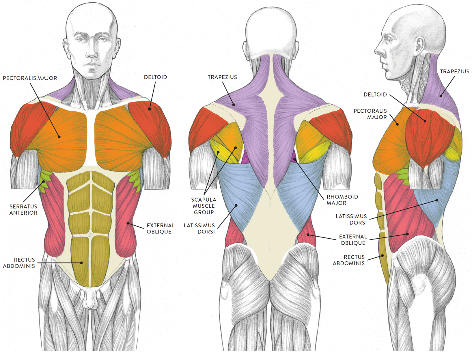
The back comprises the spine and spinal nerves, as well as several different muscle groups upper back anatomy. • raise the lateral flaps to expose parts of the latissimus dorsi muscle and define its anatomy.
Share :
Post a Comment
for "Neck And Upper Back Anatomy : How to manage neck pain with swelling in back muscles? - Dr. Kiran Sundara Murthy - YouTube"


Post a Comment for "Neck And Upper Back Anatomy : How to manage neck pain with swelling in back muscles? - Dr. Kiran Sundara Murthy - YouTube"